
Seok-Hyun (Andy) Yun, PhD
Read BioWe invent tools and techniques using light to understand biological systems better. In turn, we are curious how the biological systems can be manipulated to generate and interact with light in previously unthinkable ways. Addressing some pressing needs and chasing our intellectual curiosity, we take multidisciplinary approaches integrating physics, biology, chemistry, bioengineering, and medicine. Some of our inventions have been licensed to the industry and approved by FDA, while new technologies are being developed. Current research projects are as follows:
Bio-integrated Photonics
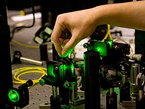
We are interested in developing a variety of biomedical photonic devices using principles, materials and structures that are biologically inspired or bio-engineered, implantable, biodegradable, wearable, and often mimicking nature at scales from nano to macro levels. Motivation of such devices is primarily for sensing, diagnostics, and therapeutics for applications in medicine, solving the limitations of conventional optical devices and conventional approaches. Often time, our work is driven by intellectual curiosity, even without specific applications in mind.
Bio lasers
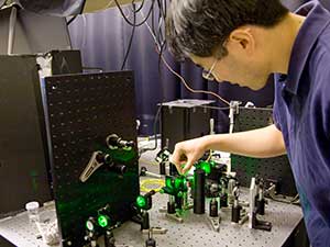
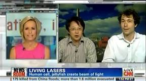
Since Theodore Maiman demonstrated the first laser a half century ago, stimulated emission and lasing have made a tremendous impact on modern science and technology. Although lasers and the coherent light they emit are omnipresent today, lasing has remained a man-made phenomenon. Generation of laser light has so far relied on artificial or engineered optical gain materials, such as doped crystals, semiconductors, synthetic dyes, and purified gases; biological materials and living organisms have not been explored as gain materials for lasers. This project introduces fluorescent proteins as a new optical gain material. The proposed research will investigate the optical amplification characteristics of biologically produced, biocompatible and biodegradable fluorescent proteins that have high quantum yield at various wavelengths across the entire visible spectrum. The fluorescent proteins offer unique properties not shared by any existing gain materials. For example, they can be expressed as functional transgene in a wide variety of organism. Intellectual merit of this project is harnessing these properties of fluorescent proteins to demonstrate novel photonic device concepts, including protein lasers in solutions and in condensed-state forms, as well as biological lasers based on single cells expressing fluorescent proteins will be explored. Successful completion of the proposed research will improve our ability to control and optimize the stimulated emission properties of fluorescent proteins and demonstrate miniature lasers and amplifiers built on the biological platform. Furthermore, it is expected to provide insights into a range of novel protein-based devices and technologies for practical applications. For example, lasing within the tissues and inside the cells may offer new possibilities of nonlinear deep imaging and intracellular sensing for bioengineering and medical diagnosis. The ability to generate laser light in vivo may enable new approaches in light-controlled therapy and drug activation.
Biocompatible and biodegradable photonics
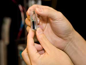
A variety of optical techniques in photomedicine require efficient delivery of light deep into and from tissues, but the limited penetration of light in tissue constitutes a serious constraint in clinical use. Fiber-optic devices or catheters have been useful in bringing a light source close to the target tissue in the body. However, delivering the light further into the tissue has remained a challenge. We are developing a new class of optical devices and previously unexplored approaches for therapy, surgery and diagnosis, as well as sensing.
In vivo Microscopy in Mice
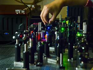
Live imaging technology represents a paradigm shift in how we study biological processes, from the traditional static 2D snapshots (i.e. histology) to dynamic, 3D view of the living specimen as processes unfold in real time. While whole-body imaging modalities such as MRI and CT have been highly successful in the clinical setting, mechanistic studies of biological processes such as disease progression and response to therapy in preclinical (animal) models require cellular details that are beyond the resolution of these whole-body imaging modalities. We have established a unique in vivocellular-level imaging resource to facilitate and accelerate collaborative biomedical research. This resource has strong imaging expertise as well as cutting-edge instrumentation that are developed in house and not commercially available. We develop and use confocal and multiphoton fluorescence imaging as well as other modalities. In particular, we have developed miniature high-resolution endomicroscopy that allows us to access internal organs minimally invasively in vivo and over time.
Brillouin Microscopy
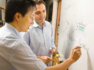
The goal of this project is to develop and validate a new biomedical imaging technology based on Brillouin light scattering for non-invasively measuring the biomechanical and rheological properties of tissues, cells, and biomaterials. Spontaneous Brillouin scattering arises from the interaction of light and sound wave that is inherently present in a sample. By detecting the spectral shifts in the scattered light, which are in the order of 10 GHz, the sample’s hypersonic viscoelastic properties can be measured at microscopic spatial resolution without physical contact. Brillouin spectroscopy has long been known in material physics and environmental sensing. However, its potential applications in biological sciences and clinical medicine have not been fully explored. We have recently developed a highly efficient spectrometer and demonstrated the possibility of in vivo Brillouin imaging. We aim to develop new practical instruments and validate them for a number of potential applications. Brillouin imaging has a potential to be a useful diagnostic tool in ophthalmology for early detection and screening of various lens and corneal problems, such as presbyopia, cataracts, and corneal ectasia. This technology may also be useful in the study of cellular biomechanics in cancer and tissue engineering.
Optical Coherence Tomography (OCT)
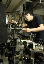
There are several projects on the development of new OCT technologies and their application to Otolaryngology, Ophthalmology, Nanomedicine, and Tissue Engineering. These projects are in part funded by NIH P41.
Group Website
For more information, please visit the Bio-Optics Lab website: http://www.intelon.org/
Selected Publications
Jiang Y, Li GY, Wang R, Feng X, Zhang Y, Yun SH
Comprehensive characterization of nonlinear viscoelastic properties of arterial tissues using guided-wave optical coherence elastography. ArXiv. 2025;:ePub - PMID: 40740519 - PMCID: PMC12310123Feng X, Li GY, Jiang Y, Shortt-Nguyen O, Yun SH
Optical Coherence Elastography Measures Mechanical Tension in the Lens and Capsule. Acta Biomater. 2025;199:252-261 - PMID: 40319993 - PMCID: PMC13196500 - DOI: 10.1016/j.actbio.2025.05.009Martino N, Yan H, Abbott G, Fahlberg M, Forward S, Kim KH, Wu Y, Zhu H, Kwok SJJ, Yun SH
Large-scale combinatorial optical barcoding of cells with laser particles. Light Sci Appl. 2025;14(1):148 - PMID: 40169572 - PMCID: PMC11962087 - DOI: 10.1038/s41377-025-01809-xCho S, Martino N, Yun SH
Half-wave nanolasers and intracellular plasmonic lasing particles. Nat Nanotechnol. 2025;20(3):404-410 - PMID: 39747602 - PMCID: PMC12125510 - DOI: 10.1038/s41565-024-01843-7- More publications ...
News
2026-10-27:
The members of the Wellman Center Career Development and Promotions Committee (CDAP), Tayyaba Hasan, Brett Bouma, Bobby Redmond, Gary Tearney and Andy Yun are pleased to announce the promotion of Nicola Martino, PhD to the rank of Instructor at Harvard Medical School effective October 1, 2026.
Dr. Martino received his PhD in Physics from Politecnico di Milano, Italy, and joined Prof. Seok-Hyun Yun's laboratory at the Wellman Center for Photomedicine in 2015. At Wellman, Dr. Martino has played a leading role in the development of laser particles (LPs), miniature semiconductor lasers that can be used as optical tags to uniquely identify and track individual cells. In a study published in Nature Photonics (2019), “Wavelength-encoded laser particles for massively multiplexed cell tagging,” he demonstrated how these particles could enable large-scale cellular identification through their distinct emission wavelengths. More recently, he led the development of combinatorial optical barcoding, in which multiple LPs are combined to generate millions of unique cellular identifiers, as reported in Light: Science & Applications (2025). These optical barcodes can be read repeatedly and non-destructively, allowing individual cells to be identified across multiple measurements and experimental platforms. LP technology has also been applied to high-dimensional flow cytometry, as demonstrated in Nature Biomedical Engineering (2024), and has contributed to the establishment of LASE Innovation Inc., a startup dedicated to its commercialization, where he serves as a scientific consultant. The work has led to two patent applications on laser particle technology.
Dr. Martino’s current research focuses on extending optical barcoding to longitudinal single-cell analysis. He is developing a new generation of particles that integrate multiple lasers into a single structure, allowing the barcode to be inherited intact through cell division. This approach enables the tracking of individual cell lineages over time and the linking of measurements from microscopy and flow cytometry with downstream single-cell sequencing. In an ongoing collaboration with Prof. Galit Lahav’s group at Harvard Medical School, he is applying these technologies to investigate how the temporal dynamics of p53 signaling relate to gene expression and treatment resistance in cancer cells. He is now developing a platform for long-term tracking and high-content profiling of individual cells, with an initial focus on the progression of cellular senescence."
Please join us in congratulating Nicola on this well-deserved promotion.
2026-09-21:
The members of the Wellman Center Career Development and Promotions Committee (CDAP) are delighted to announce the promotion of Dr. Yuxuan Jiang to the rank of Instructor at Harvard Medical School effective October 1, 2026. The CDAP membership is comprised of Tayyaba Hasan (chair), Brett Bouma, Bobby Redmond, Gary Tearney, and Andy Yun.
Dr. Jiang completed his PhD study in Mechanics at Tsinghua University in China and joined Dr. Seok-Hyun Yun’s Advanced Optics Laboratory at the MGH Wellman Center for Photomedicine. In the short term, Dr. Jiang has established a research program that integrates sophisticated wave dynamics theory with innovative optical coherence elastography (OCE) to quantitatively interrogate tissue biomechanics. He established a mechanics framework that describes shear wave dynamics in prestressed and viscoelastic biological tissues. Building on this framework, he developed an OCE technique that, for the first time, enables in situ quantification of crystalline lens stress and shear modulus, extending OCE beyond conventional stiffness characterization. The study revealed spatially heterogeneous distributions of capsular tension that are closely linked to lens fluid transport and accommodation. These works were published in International Journal of Engineering Science (2025): “Incremental dynamics of prestressed viscoelastic solids and its applications in shear wave elastography”. Acta Biomaterialia (2025): “Optical coherence elastography measures mechanical tension in the lens and capsule”.
Dr. Jiang is also extending OCE to broader applications in tissue biomechanics through a collaboration with Dr. Yanhang Zhang in the Department of Biomedical Engineering at Boston University. Playing a central role in designing and executing the project, he demonstrated in porcine aortas a stress-dependent modulation of arterial viscoelasticity, suggesting an optimizing function of arterial microstructure during cyclic deformation. This work was published in Communications Physics (2026): “Comprehensive characterization of nonlinear viscoelastic properties of arterial tissues using guided-wave optical coherence elastography”.
2025-10-17:
We are thrilled to announce that Dr. Fred Cho has been promoted to Assistant Professor. Please join us in congratulating him on this well-deserved achievement. We look forward to his continued success and the exciting contributions ahead!