


Mechanisms for light activated protein crosslinking
The proposed mechanism for crosslinking proteins begins with absorption of light energy by the dye to form a very short-lived excited singlet state, 1RB*, that rapidly converts to an excited triplet state, 3RB* (Fig. 2). The dye used for PTB, Rose Bengal (RB), strongly associates with collagen fibers in tissue. Electron transfer between the side chains of certain amino acids and a RB excited state generates charged amino acid radicals (AA•+, AA•-) and charged RB radicals (RB•+, RB•-). By reactions such as adding or releasing a proton, free radicals are formed on the collagen, e.g., AA•. Coupling of these protein radicals forms covalent crosslinks between collagen chains (Fig. 3). When the protein radicals are on the apposing sides of a wound, the covalent crosslink attaches the tissues together. These molecular “nanosutures” are the basis of the continuous attachment and formation of an immediate, strong and water-tight bond by the PTB process. When the radicals are on solubilized collagen, a collagen network is formed resulting in a gel that can encapsulate cells.
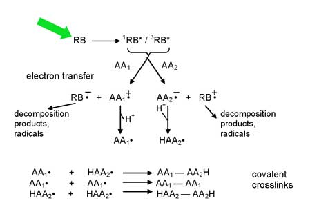 |
| Figure 2. Proposed mechanism for formation of covalent crosslinks during light-activated tissue repair. |
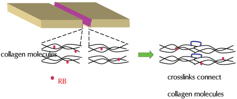 |
| Figure 3. Formation of the protein-protein covalent crosslinks (nanosutures) that attach tissues in PTB. |
Research Projects
Mechanisms for light activated protein crosslinking
Closure of surgical wounds
Cornea: sealing incisions and transplantation
Peripheral nerve repair
Tendon repair
Blood vessel repair
Neocartilage generation