


Atherosclerosis: optical diagnostics & therapy
The rupture of unstable atherosclerotic plaque frequently precedes the occurrence of myocardial infarction (MI) and stroke. The processes leading to plaque instability and the therapeutic mechanisms contributing to plaque stabilization involve a complex liaison between morphological, compositional and biomechanical factors. Some of the research areas that we are currently exploring include:
1. Intra-arterial Laser Speckle Imaging (LSI) for detecting unstable plaque
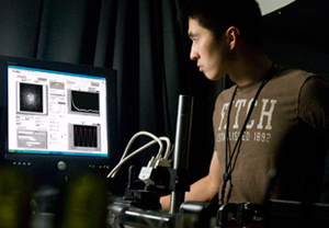
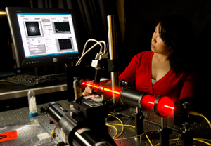
LSI is a technique we have recently developed to provide a diagnostic platform to evaluate of atherosclerotic plaque. LSI provides composite measurements related to morphological, compositional and mechanical factors, potentially yielding a powerful tool for detecting high-risk plaques. While these ex vivo studies have indicated the immense diagnostic potential of LSI, key technical challenges exist in extending LSI technology for in vivo use. We are currently developing intra-arterial LSI catheters and high-speed LSI systems to allow rapid screening of arterial segments for in vivo use in the catheterization suite.
2. Monitoring arterial mechanical properties during plaque progression
It is well established that multiple morphological, compositional and biochemical processes act cumulatively to compromise the mechanical properties of the plaque, culminating in rupture. The final event of plaque rupture is a result of the complex interplay between the viscoelastic properties of the plaque and the extrinsic luminal stresses acting upon it. Our research tests the hypothesis that during the progression of atherosclerosis, plaque viscoelastic modulus is altered and monitoring this signature metric provides a key determinant of plaque stability. We are developing new non-contact optical approaches to measure the mechanical properties of tissue and applying these techniques to monitor plaque viscoelasticity during atherosclerosis progression in animal models.
3. Develop techniques for local plaque stabilization
Another area of research in our lab investigates the development of targeted therapies to stabilize atherosclerotic plaques. This will include the development of new therapy catheters and systems, detection of new therapeutic targets, studies on the mode of cell death and its effect on plaque stabilization and regression, and the investigation of new methods to locally alter plaque composition.
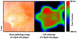 |
| The figure shows a colormap distribution of laser speckle dynamics across an atherosclerotic plaque measured by Laser Speckle Imaging (LSI). The presence of lipid rich regions of the plaque with clearly demarcated borders is evident in the LSI colormap and is corroborated in the accompanying gross pathology photograph |
| Dynamic laser speckle patterns of atherosclerotic plaques | ||
 |
 |
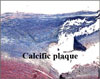 |
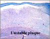
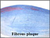
| Dynamic laser speckle images of three atherosclerotic plaques are seen here as captured using a high-speed CMOS camera. On visual inspection, stark differences in laser speckle dynamics between the lipid rich, fibrous and calcific plaque are easily observed. | ||
Measurement of soft tissue mechanics using optical techniques
In a variety of pathological processes, tissue mechanical properties are altered from the normal state. Changes in morphological, compositional and biochemical factors which characterize diseased tissue are often accompanied by alterations in the tissue mechanical properties. Optical technologies can facilitate high-resolution mapping and interrogation of tissue mechanical properties and can thus provide key information related to disease diagnosis. Our laboratory is developing optical approaches to evaluate the mechanical properties of soft tissues for a variety of applications in Cardiology, Orthopedic medicine, Otolaryngology and tissue engineering. The ability to conduct imaging via small diameter endoscopes, potentially allows the in vivo measurement of tissue mechanical properties in patients.